
About calcific tendinitis
In calcific tendinitis, chalky calcium builds up inside one of the rotator cuff tendons, most often the supraspinatus at the top of the shoulder. It is different from wear-and-tear arthritis and is most common in adults in middle age.
Deposits can sit quietly for years. Pain often flares when the body starts to reabsorb the calcium, a phase that can be intensely painful and can wake people at night. Some deposits eventually go away on their own, while others stay and keep irritating the shoulder.
Common symptoms
- Sudden, sharp shoulder pain that may come on without an injury
- Severe night pain
- Pain with lifting the arm overhead or out to the side
- A painful catch partway through arm movement
- Guarding the arm because movement hurts
Common causes and risk factors
- Changes within the tendon whose exact cause is still unclear
- Middle age, with deposits more common in women
- Possible links to diabetes and thyroid conditions
How we diagnose it
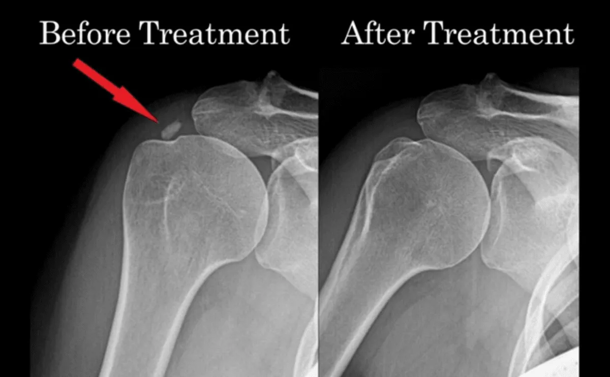
Dr. Gruner examines the shoulder and uses ultrasound at the exam table to find the deposit, measure its size, and see whether it looks hard or soft. Ultrasound also shows irritation in the nearby bursa. An X-ray confirms the calcium and helps track changes over time.
How Propel treats calcific tendinitis
Many deposits calm down with activity changes, short-term pain control, and a gradual strengthening program that keeps the shoulder moving. During a painful flare, an ultrasound-guided injection into the bursa can quiet inflammation quickly so you can sleep and move again.
When a deposit keeps causing pain, Dr. Gruner may recommend ultrasound-guided barbotage, also called lavage. Through a needle, the deposit is broken up, rinsed with fluid, and drawn out, often followed by an injection into the bursa to calm inflammation. For harder deposits or a damaged tendon, percutaneous tenotomy (Tenex) breaks up and removes calcium and damaged tendon tissue through a tiny incision so healthy tissue can regenerate. A structured rehab plan follows either procedure, and surgery is rarely needed.
What to expect at your visit
- 1.
We listen first
Your visit opens with a real conversation: how the problem started, what you have already tried, and the activity you want back.
- 2.
Exam and in-office ultrasound
A hands-on exam, then ultrasound at the exam table, often while you move, so your physician can see the tendon, joint, or nerve involved.
- 3.
A clear diagnosis and plan
You leave knowing what is wrong and what your options are, from strength-based rehab to procedures, and why we recommend each one.
- 4.
Treatment and follow-through
Most procedures happen here in the office. We coordinate with your physical therapist and check progress against the goals you set.
Rehab protocols
Dr. Gruner's week-by-week recovery guidelines. Your plan is individualized; follow these with your physician and physical therapist.
Questions about calcific tendinitis
- Will a calcium deposit in the shoulder go away on its own?
- Sometimes. Some deposits are reabsorbed by the body over months to years, often with a painful phase along the way. Others stay and keep causing symptoms, which is when a procedure may help.
- Is calcific tendinitis the same as arthritis?
- No. Calcific tendinitis is calcium inside a tendon, while arthritis is wear of the joint cartilage. They can cause similar pain, which is why an exam and imaging matter.
- How is Tenex used for calcific tendinitis?
- Under live ultrasound, a small probe is guided into the deposit through a tiny incision. It breaks up and removes the calcium along with damaged tendon tissue. Most people return to light daily use of the arm soon after and then begin rehab.